Successful Management of Gastric Leiomyoma
Dogs

Patient: Edie, 13-year-old Maltese mix
Presenting Concern: Incidental gastric mass identified on ultrasound during pre-dental screening
Referral Reason: Further diagnostics and surgical consultation
Diagnostics & Workup:
Edie was initially evaluated at her primary care clinic following elevated liver values and mild anemia. During pre-dental ultrasound, a 4 cm hypoechoic lobulated gastric mass was identified at the cardia region, partially protruding into the gastric lumen. Thoracic radiographs and abdominal ultrasound revealed no evidence of metastasis but noted mild cardiomegaly, hepatomegaly, renal mineralization, and bilateral adrenal thickening.
She was referred to AVES for advanced staging, including:
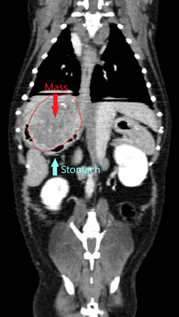
CT thorax/abdomen: Confirmed a partially mineralized gastric mass (approx. 4.1 x 3.8 x 3.2 cm) near the gastroesophageal junction. No metastatic disease observed.
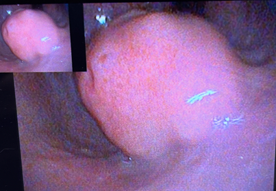
Upper GI endoscopy with biopsies: The mass appeared pedunculated with intact mucosal surface; biopsies suggested hyperplastic polyp with chronic lymphoplasmacytic gastritis and Helicobacter-like organisms; however, due to inconclusive findings, surgery was recommended for definitive diagnosis and treatment.
Surgical Management:
A partial gastrectomy was performed, carefully excising the mass with narrow margins while preserving the lower esophageal sphincter. Intraoperative recovery was initially uneventful, although Edie did experience transient post-operative upper airway obstruction likely secondary to transient laryngeal dysfunction; she responded well to oxygen supplementation and supportive care.
Histopathology:
Final histopathology revealed a spindle cell neoplasm most consistent with a benign leiomyoma. Margins were clear (closest at 1.4 mm), and no mitotic activity suggesting malignancy was identified. Immunohistochemistry was offered to further differentiate from GIST, but clinical course supported benign behavior.
Outcome:
Edie recovered fully post-operatively, resumed normal appetite, and has shown no recurrence or clinical concerns. Her prognosis is excellent with full expected life expectancy.
Key Takeaway:
This case highlights the importance of multimodal diagnostics, thorough staging, and careful surgical planning in managing complex gastric masses. Thanks to early detection, timely referral, and collaborative care, Edie has returned to a full, happy life.